Private Scans
Ultrasound scans that get you the answers you want on the same day!
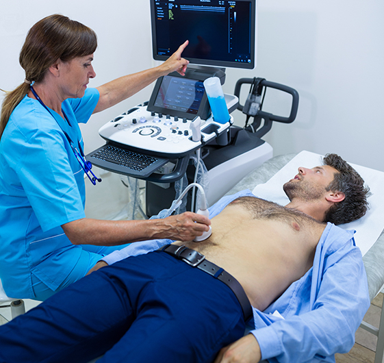
If your GP has referred you for an ultrasound scan and the NHS wait is just to long, don’t worry, you can self refer to us for your scan usually the same day or the same week. Book a private ultrasound scan today with a qualified MSK specialist radiographers or sonographers at one of our private clinics or diagnostic centres and receive your results back the same day. All MSK scan are just £120 fully inclusive with the report for your GP sent the same day.
Soft Tissue Lumps & Bumps
Firstly, most long-standing lumps and bumps are harmless. However, many people are concerned about the nature of the lump, especially if this starts to change in size or has become symptomatic (painful, sore, tender, redness, bruising)
Ultrasound is widely accepted as the first-line diagnostic tool in the assessment of palpable soft tissue lumps and bumps and is able to assess the size, shape, internal characteristics and more often than not, the origin of the lump.
This type of scan does not require any specific preparation and can quickly evaluate the affected area. Most common lumps and bumps include lipomas (fatty lumps), ganglions, cysts, effusions (fluid), tendon thickening, fibromas, neuromas and nodules.
An ultrasound scan is most often the easiest way to assess lumps and bumps of the soft tissues.
The purpose is to examine most parts of the body (excluding breast tissue). This includes assessment of the superficial tissues (skin layers and underlying muscles).
Common examples for having this ultrasound scan are to investigate the causes of soft tissue swellings and lumps for conditions such as ganglions, lipomas, cysts, skin changes, foreign bodies, hernias and unexplained swellings in most parts of the body.


Shoulder & Back
Shoulder pathology is the third most common musculoskeletal disorder after back and neck pain.
Ultrasound of the shoulder (also referred to as the rotator cuff) is an excellent imaging modality for assessment of the tendons, ligaments, muscles and the associated soft connective tissues.
If you are experiencing pain, restricted movement of the joint or pain following an injury, musculoskeletal ultrasound is able to evaluate these structures with a comparable accuracy to a MRI scan.
Ultrasound has the added benefit of being a dynamic examination so that the shoulder and its mechanics can be evaluated during real-time movement
Common examples for having this ultrasound scan are to investigate the causes of shoulder pain, possible rotator cuff injury or tears, subacromial impingement, bursitis, calcific tendinosis and degenerative changes such as arthritis.
Elbow and Forearm
The most common causes of pain in the elbow are is due to overuse injuries, often associated with repetitive movements. Overuse injuries of the elbow result in either medial or lateral tendon inflammation or damage, otherwise known as tennis or golfers elbow, or lateral epicondylitis and medial epicondylitis.
However, it is not only repetitive sport movements that cause these injuries with many recurring day-to-day mechanical movements resulting in the same symptoms.
Ultrasound can also evaluate the bursae of the elbow small fluid sacs that help prevent friction, nerves, bony surfaces, ligaments, tendons and musculature. Please also see wrist and hand ultrasound scan if you have symptoms in those areas
The purpose is to examine the elbow for a range of conditions. This includes assessment of the joint, the flexor and extensor tendons, muscles, ligaments, bursa and soft-tissue swellings.
Common examples for having this ultrasound scan are to investigate the causes of elbow pain, possible injury or inflammation of the tendons for conditions such as tennis and golfers elbow, olecranon bursitis, tendinosis and degenerative changes such as arthritis.


Hand & Wrist
Around 26% of the UK population suffer from wrist and hand pain.
There are many reasons why wrist and hand pain may occur, just some of the causes include; carpal tunnel syndrome, synovitis, arthritis, tendinopathy, ligament damage, trigger finger, tendon contracture, soft tissue or tendon lesions and soft tissue lumps and bumps such as ganglions and fluid within the tendon sheaths.
Ultrasound is able to assess these conditions with a high degree of accuracy.
The purpose of this the ultrasound scan is to examine and assess the wrist joint and the carpal tunnel, the flexor and extensor tendons, ligaments, small joints of the hand, associated annular and cruciate pulleys and soft-tissue swellings.
Common examples for having this ultrasound scan are to investigate the causes of wrist and hand pain, possible injury or inflammation of the tendons for conditions such as carpal tunnel syndrome, trigger finger, ganglions, tendinosis and degenerative changes such as arthritis.
Hip, Groin and upper thigh
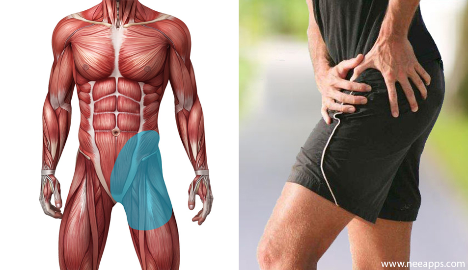
Hip Pain can affect both younger and older adults for various reasons.
In the younger adult, this may be due to a sporting muscle injury, laxity of the tendons or joint, nerve irritation, snapping hip, bursitis and ligament strains. Degenerative and arthritic changes are more common, but not exclusive, in older adults. While labral tears, hip effusions and bursitis are common to all age groups.
Ultrasound proves to be a good diagnostic tool in the assessment of the hip joint, surrounding connective and soft tissues.
The purpose is to examine the hip joint and upper thigh for a range of conditions. This includes assessment of the joint, the adductor tendons, muscles, ligaments, iliopsoas and trochanteric bursae and the hip joint.
Common examples for having this ultrasound scan are to investigate the causes of hip pain, possible injury or inflammation of the tendons for conditions such as bursitis, tendinosis, degenerative changes such as arthritis, hip effusions and labral tears.


Knee
Knee problems are very common occurring in people of all ages.
This can interfere with daily activities from taking part in sporting activities to getting up from a chair and walking.
The knee is a complex joint made up of bone, cartilage, ligaments, tendons and muscles, all prone to injury and degenerative changes. As well as injury, certain types of arthritis can lead to cartilage and bone destruction, giving the symptoms of pain, swelling and a loss of flexibility within the affected joint.
Ultrasound is a well-established imaging method for assessing these structures and the many associated pathological processes.
The purpose is to examine the knee joint for a range of conditions. This includes assessment of the joint, the tendons, associated muscles, lateral and medial collateral ligaments, bursae, the lateral and medial menisci and the posterior knee.
Common examples for having this ultrasound scan are to investigate the causes of knee pain, possible injury or inflammation of the tendons and ligaments, for conditions such as bursitis, tendinosis, degenerative changes such as arthritis, knee effusions and meniscal cysts / degeneration, cysts in the posterior knee and
Ankle & Foot
The purpose is to examine the ankle & foot for a range of conditions. This includes assessment of the ankle joint, the anterior, medial and lateral tendons, the Achilles tendon, associated soft tissues, musculature, bursae and small joints of the ankle and foot.
Common examples for having this ultrasound scan are to investigate the causes of ankle & foot pain, possible injury or inflammation of the tendons, for conditions such as bursitis, tendinosis, degenerative changes such as arthritis, joint effusions and ganglions / tendon and ligament tears, neuromas, fibromas and plantar fasciitis.
A unique advantage of ultrasound is the ability to assess the ankle and foot dynamically in real time, revealing abnormalities that may be inadequately visualised on static images. It is also easy to compare the affected area with the non-symptomatic opposite side.
Pain in the ankle and foot can often be difficult to pinpoint to a specific area, but ultrasound is able to evaluate much of the anatomy within the ankle and foot.
Common problems visualised with ultrasound include; soft tissue masses, Morton’s neuroma, bursitis or capsulitis of the joints, ligament injuries, tendinopathy and tears, heel spurs and plantar fasciitis and tarsal tunnel syndrome. These are just some of the conditions we are able to detect.


Hernia
An abdominal wall hernia is when an organ or body tissue squeezes through a weak spot in the wall of the abdomen, often causing a lump which you can feel and may come and go.
Hernias can occur in a number of different places throughout the abdomen, particularly if you have had previous surgery (an incisional hernia).
Groin hernias are up to 10 times more common in men, and this is when the internal body fat or part of the bowel can pass through a weak spot into the inguinal canal – a pathways for structures pass from the abdomen to the genitalia.
The most common types of hernia are:
- Inguinal (inner groin)
- Incisional (at the site of previous surgery)
- Femoral (outer groin)
- Umbilical (belly button)
Hernias can occasionally cause pain or discomfort and often the lump you feel comes and goes, particularly when you strain or cough but can also get stuck – known as an incarcerated hernia – where tissues get trapped in the outpouching.