ALL OUR PELVIC SCANS ARE PERFORMED BY ONLY FEMALE SONOGRAPHERS
A pelvic ultrasound scan allows your Sonographer to see your bladder, cervix, uterus, fallopian tubes, and ovaries.
The test is done in two ways:
Transabdominal
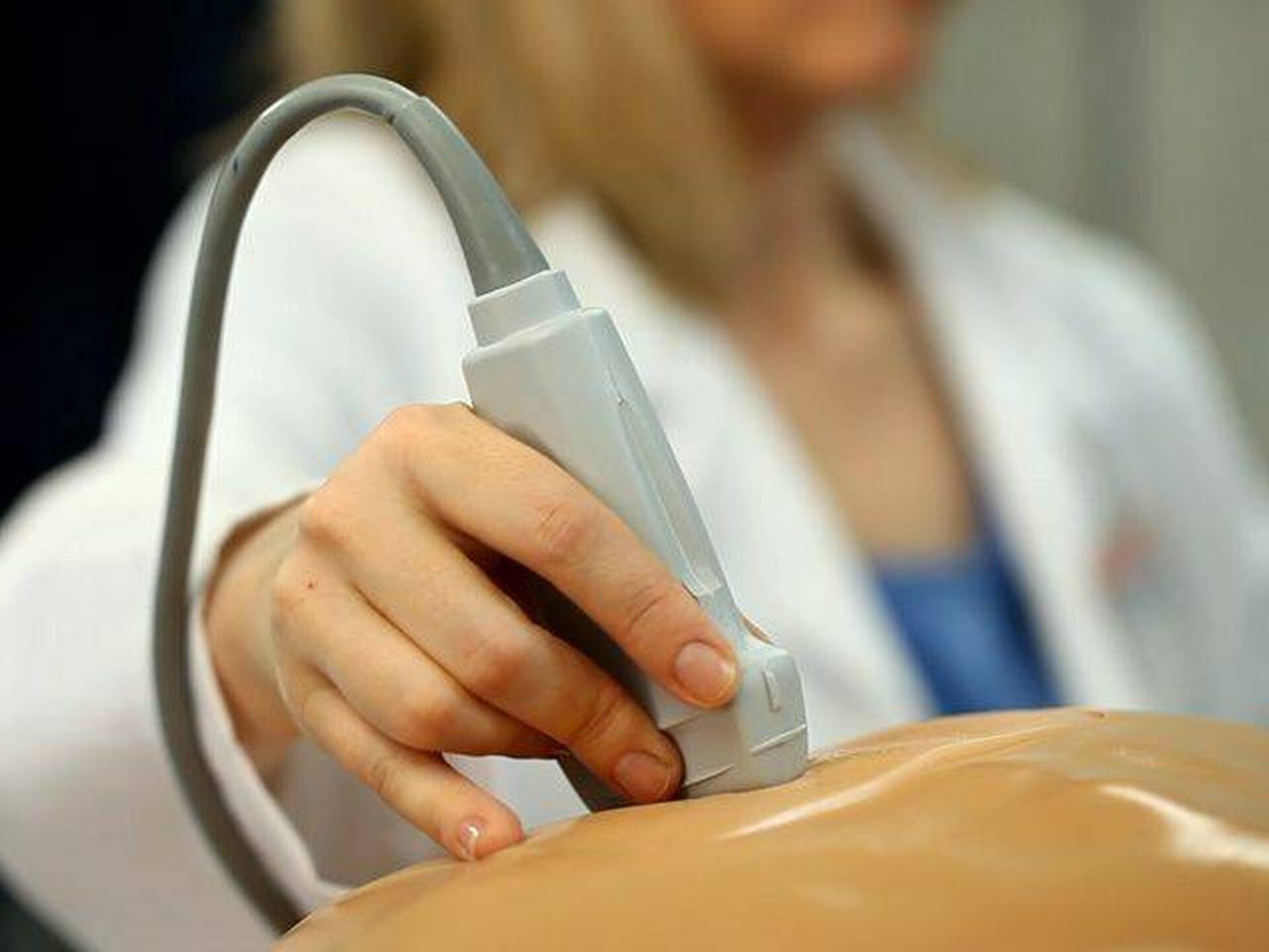
- A simple scan where the scan probe gently glides across your lower tummy region.
Transvaginal
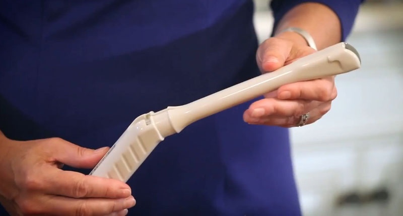
- A thin, lubricated transducer is placed inside the vagina. This does not hurt and should not even be uncomfortable. Our highly trained female specialist staff have many years experience in performing these scan.
A pelvic ultrasound test is done for the following reasons:
- Find the cause of urinary problems.
- Find out what’s causing pelvic pain.
- Look for causes of vaginal bleeding and menstrual problems.
- Check for growths or masses like ovarian cysts or uterine fibroids.
- Look for the correct placement of an intrauterine device (IUD).
These are the most common reasons, but there may be other reasons for your doctor to recommend a pelvic ultrasound.


How can you prepare for your pelvic exam?
If you have transabdominal ultrasound, you must drink 4 to 6 glasses of water about an hour before the test. This will fill your bladder and make for a clearer picture.
If you are having both transabdominal and transvaginal ultrasounds done, you’ll start with a full bladder. You will be asked to empty it before the transvaginal ultrasound.
What happens during your pelvic ultrasound exam?
For a transabdominal ultrasound
You will lie on your back on an examination table. A gel-like substance will be applied to your abdomen.
The transducer will be pressed against the skin and moved around over the area being studied.
Images of structures will be displayed on the computer screen. Images will be recorded on various media for the health care record.
Once the procedure has been completed, the gel will be removed.
You may empty your bladder when the procedure is completed.
For a transvaginal ultrasound
If asked to remove clothing, you will be given a gown to wear.
You will lie on an examination table, with your feet and legs supported as for a pelvic examination.
A long, thin transvaginal transducer will be covered with a plastic or latex sheath and lubricated. The tip of the transducer will be inserted into your vagina. This may be slightly uncomfortable.
The transducer will be gently turned and angled to bring the areas for study into focus. You may feel mild pressure as the transducer is moved.
Images of organs and structures will be displayed on the computer screen. Images may be recorded on various media for the health care record.
Once the procedure has been completed, the transducer will be removed.


What else should you know about the exam?
- With a transabdominal ultrasound, you will feel light pressure from the transducer as it passes over your belly. If you have an injury or
pelvic pain, the pressure may be uncomfortable but painful and the clinician will check on you for pain throughout the scan. - With a transvaginal ultrasound, the transducer probe is gently put into your vagina. If at any time
you become uncomfortable do let the sonographer know and they will stop the scan. - You will not hear or feel the sound waves.
What happens after the exam?
- There is no particular type of care required after a pelvic ultrasound. You may resume your regular diet and daily activities unless your doctor advises you differently.
- There are no confirmed adverse biological effects on patients or instrument operators caused by exposures to ultrasound at the intensity levels used in diagnostic ultrasound.
- But depending on your particular results, the doctor may give you additional or alternative instructions after the procedure.
